DOC_AGCK_PIF_3
| Accession: | |
|---|---|
| Functional site class: | AGC Kinase docking motif |
| Functional site description: | The AGC kinases constitute a large family of serine/threonine protein kinases consisting of 60 members, including the cAMP- and cGMP-dependent protein kinases (PKA and PKG), the protein kinase C family (PKC), PKB/Akt, ribosomal protein S6 kinases, and the 3-phosphoinositide-dependent protein kinase (PDK1). They regulate many critical processes including metabolism and cell proliferation. Members of this family contain a hydrophobic surface in the N-terminal lobe of their catalytic domain, called the PDK1 Interacting Fragment (PIF) pocket, and a non-catalytic C-terminal tail containing different motifs, including the AGCK docking motif that interacts with the PIF pocket. Both these regions are conserved in Eukaryotic AGC kinases, except for PDK1, which lacks the C-tail. The AGCK docking motif mediates intramolecular interactions to the PIF pocket, serving as a cis-activating module, but can also act as a PDK1 docking site that trans-activates PDK1, which in turn will phosphorylate the docked AGC kinase. |
| ELMs with same func. site: | DOC_AGCK_PIF_1 DOC_AGCK_PIF_2 DOC_AGCK_PIF_3 |
| ELM Description: | The AGCK docking motif of some AGC kinases, including PKA, does not contain a phosphorylatable serine/threonine residue or an acidic aspartate/glutamate residue present in the DOC_AGCK_PIF_1 and DOC_AGCK_PIF_2 motif variants, respectively. Instead, this variant only consists of the first two core aromatic residues preceding this site in the other motif variants, and in this case the latter of these two aromatic residues is located at the C-terminal position of the kinase. So far, the two aromatic residues have only been observed to be phenylalanines. Mutation or deletion of these residues greatly diminishes PKA activity and PKA-PDK1 interaction. |
| Pattern: | F..F$ |
| Pattern Probability: | 6.030e-07 |
| Present in taxon: | Eukaryota |
| Interaction Domain: |
Pkinase (PF00069)
Protein kinase domain
(Stochiometry: 1 : 1)
|
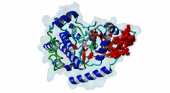
This entry covers an auto-activating linear motif of AGC group kinases. Several variants of the motif exist, and for many kinases, the motif has been shown to operate in trans to bind and activate the upstream activating kinase PDK1. To make matters more complicated, some variants are regulated by phosphorylation. The AGC kinases regulate critical processes including metabolism, cell growth, proliferation, survival and differentiation, hence deregulation of these enzymes is a causative factor in different diseases such as cancer and diabetes. Solved structures of AGC kinases show the typical bilobal kinase fold of the kinase domain, consisting of a small N-terminal lobe (N-lobe) and a larger C-terminal lobe (C-lobe). Regulation of kinase activity is mainly achieved through phosphorylation of the activation or T-loop, located in the C-lobe and connected to the N-lobe through the alpha-C helix. This modification results in conformational changes, mainly in the alpha-C helix, that reposition key catalytic and substrate binding residues. Sandwiched between the N- and C-lobe is an ATP-binding site that provides the phosphate-donor during phosphorylation. Repositioning of the alpha-C helix upon kinase activation allows formation of a salt bridge between an alpha-C helix glutamate and a conserved lysine residue within the beta-3 strand that interacts with the alpha and beta phosphates of ATP (Pearce,2010). The non-catalytic C-terminal tail of the kinase is also involved in repositioning of the alpha-C helix. The alpha-C helix is part of a hydrophobic pocket and an adjacent phosphate-binding site in the N-lobe, called the PIF pocket, which interacts with the AGCK docking motif (PDK1 Interacting Fragment (PIF) / hydrophobic motif (HM)) that is present in the C-tail of AGC kinases. This interaction stabilizes the active conformation of the alpha-C helix through an allosteric mechanism. Both the PIF pocket and the C-terminal region are conserved in Eukaryotic AGC kinases, except for PDK1, which lacks the C-terminal part. The AGCK docking motif mediates intramolecular interactions to the PIF pocket, serving as a cis-activating module, together with other regulatory sequences present in the C-tail of the kinase. However, in some kinases it also serves as a PDK1 docking site that trans-activates PDK1, which itself does not possess this regulatory region. Activated PDK1 in turn will phosphorylate and activate the docked AGC kinase (Mora,2004). Several AGC kinases are involved in mediating signaling downstream of phosphatidyl-inositol-4,5-bisphosphate 3-kinase (PI3K) in response to a wide range of stimuli such as growth factors and hormones. PDK1 functions as a common upstream activator by phosphorylating the other AGC kinases at their activation loop. The PDK1 PIF pocket serves both as an allosteric regulatory site for PDK1 activity and as a docking site for the AGC kinases it phosphorylates, by binding to the AGCK docking motif that is present in the C-tail of its substrates. AGC kinases in their inactive state have an incompatible PIF pocket due to the alpha-C helix being disordered, meaning that their AGCK docking motif is available for binding to the PIF pocket of PDK1, which becomes trans-activated. After being phosphorylated by PDK1, the AGCK docking motif can engage in an intramolecular interaction with its functional PIF pocket, resulting in release from PDK1 and full activation of the kinase. The AGCK docking motif generally appears as three aromatic residues, most often phenylalanines, surrounding a phosphorylatable serine or threonine residue (DOC_AGCK_PIF_1). Phosphorylation of this serine/threonine increases the affinity of the motif for the PIF pocket, which allows fine-tuning the cis and trans interactions of the motif. The mechanisms and kinases involved in phosphorylation of the AGCK docking motif differ for the different kinases. Some alternative patterns of the motif exist. In atypical PKC forms and PKC-like (PKN) kinases, the phosphorylatable serine or threonine residue is replaced by an acidic phosphomimetic aspartate or glutamate residue (DOC_AGCK_PIF_2). In other AGC kinases, including PKA, the motif is located at the very C-terminal and contains only the first two core aromatic residues (DOC_AGCK_PIF_3). In many cases, full activation of the AGC kinases is also dependent on additional signals that are specific for each kinase and that provide spatial and conformational regulation. |
-
A 3-phosphoinositide-dependent protein kinase-1 (PDK1) docking site is
required for the phosphorylation of protein kinase Czeta (PKCzeta ) and
PKC-related kinase 2 by PDK1.
Balendran A, Biondi RM, Cheung PC, Casamayor A, Deak M, Alessi DR
J Biol Chem 2000 Jul 7; 275 (27), 20806-13
PMID: 10764742
-
The PIF-binding pocket in PDK1 is essential for activation of S6K and SGK,
but not PKB.
Biondi RM, Kieloch A, Currie RA, Deak M, Alessi DR
EMBO J 2001 Aug 15; 20 (16), 4380-90
PMID: 11500365
-
High resolution crystal structure of the human PDK1 catalytic domain
defines the regulatory phosphopeptide docking site.
Biondi RM, Komander D, Thomas CC, Lizcano JM, Deak M, Alessi DR, van Aalten DM
EMBO J 2002 Aug 15; 21 (16), 4219-28
PMID: 12169624
-
A phosphoserine/threonine-binding pocket in AGC kinases and PDK1 mediates
activation by hydrophobic motif phosphorylation.
Frodin M, Antal TL, Dummler BA, Jensen CJ, Deak M, Gammeltoft S, Biondi RM
EMBO J 2002 Oct 15; 21 (20), 5396-407
PMID: 12374740
-
Crystal structure of an activated Akt/protein kinase B ternary complex
with GSK3-peptide and AMP-PNP.
Yang J, Cron P, Good VM, Thompson V, Hemmings BA, Barford D
Nat Struct Biol 2002 Dec; 9 (12), 940-4
PMID: 12434148
-
Growth signalling pathways in Arabidopsis and the AGC protein kinases.
Bogre L, Okresz L, Henriques R, Anthony RG
Trends Plant Sci 2003 Sep; 8 (9), 424-31
PMID: 13678909
-
Phosphoinositide-dependent protein kinase 1, a sensor of protein
conformation.
Biondi RM
Trends Biochem Sci 2004 Mar; 29 (3), 136-42
PMID: 15003271
-
PDK1, the master regulator of AGC kinase signal transduction.
Mora A, Komander D, van Aalten DM, Alessi DR
Semin Cell Dev Biol 2004 Apr; 15 (2), 161-70
PMID: 15209375
-
In vivo activation of protein kinase A in Schizosaccharomyces pombe
requires threonine phosphorylation at its activation loop and is dependent
on PDK1.
Tang Y, McLeod M
Genetics 2004 Dec; 168 (4), 1843-53
PMID: 15611161
-
In vivo role of the phosphate groove of PDK1 defined by knockin mutation.
Collins BJ, Deak M, Murray-Tait V, Storey KG, Alessi DR
J Cell Sci 2005 Nov 1; 118, 5023-34
PMID: 16219676
-
Regulation of the interaction between protein kinase C-related protein
kinase 2 (PRK2) and its upstream kinase, 3-phosphoinositide-dependent
protein kinase 1 (PDK1).
Dettori R, Sonzogni S, Meyer L, Lopez-Garcia LA, Morrice NA, Zeuzem S, Engel M, Piiper A, Neimanis S, Frodin M, Biondi RM
J Biol Chem 2009 Oct 30; 284 (44), 30318-27
PMID: 19723632
-
The nuts and bolts of AGC protein kinases.
Pearce LR, Komander D, Alessi DR
Nat Rev Mol Cell Biol 2010 Jan; 11 (1), 9-22
PMID: 20027184
(click table headers for sorting; Notes column: =Number of Switches, =Number of Interactions)
| Acc., Gene-, Name | Start | End | Subsequence | Logic | #Ev. | Organism | Notes |
|---|---|---|---|---|---|---|---|
| Q9LSF1 OXI1 OXI1_ARATH |
418 | 421 | WVKGLNNNHDLESDNNFLVF | TP | 5 | Arabidopsis thaliana (Thale cress) | |
| Q5I6E9 Adi3 Q5I6E9_SOLLC |
697 | 700 | KRVVGADAKSGGKYLDFEFF | TP | 3 | Solanum lycopersicum (Tomato) | |
| P06244 TPK1 KAPA_YEAST |
394 | 397 | EDINYGVQGEDPYADLFRDF | TP | 3 | Saccharomyces cerevisiae S288c | |
| P05132 Prkaca KAPCA_MOUSE |
348 | 351 | EEEEIRVSINEKCGKEFTEF | TP | 3 | Mus musculus (House mouse) | |
| P17612 PRKACA KAPCA_HUMAN |
348 | 351 | EEEEIRVSINEKCGKEFSEF | TP | 2 | Homo sapiens (Human) |
Please cite:
ELM-the Eukaryotic Linear Motif resource-2024 update.
(PMID:37962385)
ELM data can be downloaded & distributed for non-commercial use according to the ELM Software License Agreement
ELM data can be downloaded & distributed for non-commercial use according to the ELM Software License Agreement