| Accession: | |
|---|---|
| Functional site class: | Phosphotyrosine ligands bound by SH2 domains |
| Functional site description: | Src Homology 2 (SH2) domains are small modular domains found within a great number of proteins involved in different signalling pathways. They are able to bind specific motifs containing a phosphorylated tyrosine residue, propagating the signal downstream by promoting protein-protein interactions and/or modifying enzymatic activities. Different families of SH2 domains may have different binding specificity, which is usually determined by a few residues C-terminal with respect to the pY (positions +1 to +4). Non-phosphorylated peptides do not bind to the SH2 domains. Several different binding motifs are known, for example: pYEEI (Src-family SH2 domains), pY [IV].[VILP] (SH-PTP2, phospholipase C-gamma), pY.[N] (GRB2). The interaction between SH2 domains and their substrates is however dependent also on cooperative contacts of other surface regions. |
| ELMs with same func. site: | LIG_SH2_CRK LIG_SH2_GRB2like LIG_SH2_NCK_1 LIG_SH2_PTP2 LIG_SH2_SFK_2 LIG_SH2_SFK_CTail_3 LIG_SH2_STAP1 LIG_SH2_STAT3 LIG_SH2_STAT5 LIG_SH2_STAT6 |
| ELM Description: | Class IC SH2 domains bind a pTyr motif with specificity for Asn at the pY+2 position. Peptide scanning experiments define two main preferences at the pY+1 position that include acidic or hydrophobic residues (Tinti,2013; Huang,2008). While Grb2/Grb7/Grap/Gads Sh2s bind to both types of motifs, Grb10/14 and TNS1/4 have a strong preference for acidic residues at pY+1. At pY+1, hydrophobic residues make Van der Waals interactions with W121, F108 and Q106 of the SH2 domain, while acidic residues extend further into the groove, making polar interactions with Q106 and N143. The Gads SH2 domain has a more selective pY+1 pocket which disfavours bulky or large aliphatic residues (Cho,2004). The pTyr provides the basal affinity, binding into a highly conserved pocket through multiple stabilizing interactions. The binding mode is determined by the conformation of the EF and BG loops, which plug access to the pY+3 pocket in these SH2 domains forcing the ligand to adopt a closed Type I β-turn conformation that exposes the +2 Asn prominently for binding (Kaneko,2010; Rahuel,1996). The Asn residue H bonds with the backbone of residues K109 and L120 in the SH2 domain. Proline is excluded from position pY+1 as it would prevent the U-shaped turn conformation (Huang,2008; Liu,2010). More open or twisted turn motifs are present in AICD, CDC28 and ErbB2 (Das,2011; Inaba,2017; Ivancic,2003). The peptide ligand is held in place by backbone H bonds of pY-1 and pY+1 to the SH2 domain, and by an intramolecular H bond between pY+3 and pTyr that stabilizes the turn. Val or Met at pY+3 can make weak hydrophobic interactions with the SH2 domain. Positive charge in pY+3, pY+4 and pY+5 provides specificity, preventing binding to Class 1C SH2s with a positive surface patch surrounding the pTyr pocket (Grb2) but favouring Sh2s with negatively charged surfaces (Grb7) (Fiddes,1998; Spuches,2007). Grb7,10,14 and TNS1,4 have reduced specificity for N at +2 and can bind motifs not matched by the ELM pattern. |
| Pattern: | (Y)([EDST]|[MLIVAFYHQW])N. |
| Pattern Probability: | 0.0003175 |
| Present in taxon: | Metazoa |
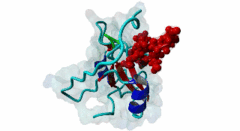
| Interaction Domain: |
SH2 (PF00017)
SH2 domain
(Stochiometry: 1 : 1)
|
The Src Homology 2 (SH2) domain is a major protein interaction module that is central to tyrosine kinase signaling. Over 120 SH2 domains are predicted in the human genome (Liu,2011). Among SH2 domain-containing proteins are kinases, phosphatases adaptors, ubiquitin ligases, transcription factors, guanine nucleotide exchange factors. The many processes involving SH2 domains range from mitogenic signaling to T cell activation. Mutations identified in many SH2 domain-containing proteins as well as the SH2 domain itself are associated with human diseases ranging from cancers, diabetes, to immunodeficiencies. SH2 domains are phosphotyrosine recognition domains, often mediating transient interactions with target proteins. The binding affinity of an SH2 domain to a pTyr containing ligand is moderate, with the typical affinity range between 0.1 µМ to 10 µМ for equilibrium dissociation constant values (Kd) (Kaneko,2012). The structure of the SH2 domain consists of a central antiparallel β-sheet formed by three or four β strands flanked by two α helices. In the canonical mode of SH2 binding, regions on either side of the central β sheet are involved in ligand binding. The N-terminal region is most conserved and contains the pTyr binding pocket. The C-terminal half of the SH2 domain exhibits greater structural variability and provides a platform for accommodating different kinds of SH2-binding motifs. Three loops surround the peptide binding pocket and are important for specificity: Because these loops can be flexible, considerable variation in peptide binding can apply for any given SH2 domain. For the majority of experimentally solved SH2:peptide ligand complex structures, the bound pTyr peptide forms an extended conformation and binds perpendicularly to the central β strands of the SH2 domain. However motifs that form alternative conformations are also identified as in the case of the GRB2 SH2 domain binding motif (Nioche,2002) where the motif forms a β-turn upon binding. Grb2 is a good example of a bifunctional adaptor protein that brings proteins into close proximity, allowing signal transduction through proteins that can span different compartments. SPOT arrays provide an overview of different SH2 specificities (Huang,2008) although it is clear that they do not fully capture all the possible motifs for any given SH2. SH2s fall into groups with related specificities such as the GRB2-like set with a preference for YxN, the Src-like family with a preference for Y--# or the unique Stat3 YxxQ preference. SPOT arrays indicate that some SH2s might have quite poor specificity, for example PLCγ1_C and GRB7: These may be quite promiscuous. A large set of SH2 motif patterns has been made available, based on the SPOT arrays and other available data [Samano-Sanchez,2023]. Because of overlapping specificities amongst SH2 domains, it is unlikely to be clear which proteins bind to a new pTyr candidate SH2-binding motif. Therefore temporal and spatial colocalization should be evaluated and ultimately direct in-cell binding demonstrated as well as interaction affinities measured by in vitro binding assays. In addition, some motifs might be bound by multiple SH2s, for example as part of a sequential signaling process. |
(click table headers for sorting; Notes column: =Number of Switches, =Number of Interactions)
Please cite:
ELM-the Eukaryotic Linear Motif resource-2024 update.
(PMID:37962385)
ELM data can be downloaded & distributed for non-commercial use according to the ELM Software License Agreement
ELM data can be downloaded & distributed for non-commercial use according to the ELM Software License Agreement